
Uncovering Concussion Clues
November 22, 2017
Findings from a recent Western-led survey may indicate that young athletes who suffer concussions may be returning to the field, court or ice too soon, as their brains are continuing to change long after they are cleared for action.
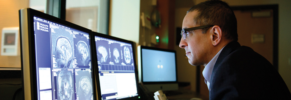
Western researcher Ravi Menon and his team at Robarts Research Institute and Schulich School of Medicine & Dentistry have shown that young hockey players who have suffered concussions still show changes in the white matter of the brain months after being cleared to return to play. The findings were published in this month’s issue of Neurology, the medical journal of the American Academy of Neurology.
The study looked at MRI brain scans from 17 Bantam-level hockey players between the ages of 11-14, who suffered a concussion during the regular season. Most of the concussions were a result of falls that resulted in a hit to the back of the head.
The athletes had their brains scanned within 24-72 hours of the initial concussion and again three months post-concussion at Western’s Centre for Functional and Metabolic Mapping. At the time of the three-month scan, all the players reported no symptoms on clinical evaluations and were cleared to return to the ice following the standard concussion consensus return-to-play protocol.
“What the MRI shows is there are still changes occurring in the brain even after the clinical tests have returned to normal,” Menon explained. “This is potentially of some concern and we’d like to understand this further to determine if these are normal healthy changes or if they are indicative of something that might be going wrong.”
The advanced MRI data was analyzed by PhD Candidate Kathryn Manning. She and her team looked at diffusion, functional and spectroscopy MRI data. On both the acute and the three-month scans, Manning said they saw damage in the very long fibre tracks in the brains of the concussed players and also saw ‘hyper-connectivity’ in some areas of the brain, which suggests the brain may still have been trying to compensate for the injury.
“We saw there were prolonged abnormalities in terms of the white matter in the brain,” said Manning, noting these changes are only visible using high field-strength MRI and these sophisticated analytical methods. “On a normal clinical MRI scan, you typically see the structural images of the brain, and for a mild brain injury like a concussion, we aren’t able to see the underlying changes we were able to see using these advanced methods.”
For Schulich professor Dr. Lisa Fischer, the news is promising for concussion diagnosis. She sees hundreds of concussed athletes each year at the Fowler Kennedy Sport Medicine Clinic at Western. Currently, the clinical protocols used to diagnose concussions and evaluate return-to-play are based mainly on subjective reporting from the patient.
“If we can come up with a clinically relevant, objective measure for concussion diagnosis and recovery, we can make safer decisions about return to play,” she said. “This study has the potential to help develop that.”


