Imaging – Did you know?...
April 18, 2016

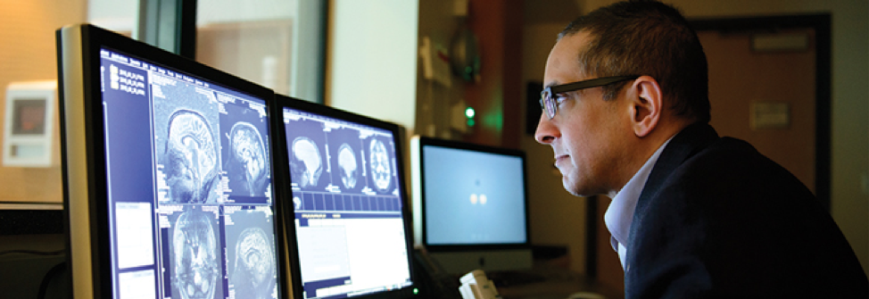
At Robarts Research Institute, advancements in imaging are enabling scientists to see inside the human body with incredible precision and detail.
Robarts houses Canada’s most robust and sophisticated imaging facilities, including cutting-edge MRI suites, CT scanners, ultrasound machines, and cellular and molecular imaging technologies. With access to these facilities, our scientists are focused on improving the understanding, early diagnosis and treatment of human disease through the development of innovative techniques and instrumentation.
Our groundbreaking imaging research supports a wide range of conditions affecting human health, including cancer, neurodegenrative disorders, lung diseases, bone and joint diseases and heart disease.
Robarts imaging scientists are national and international leaders in their fields, and excel in interdisciplinary and collaborative research that is focused on fostering creative solutions to human disease. With a patient-centred perspective, together we are striving for a healthier future.
From the bench to the bedside: Our imaging research is changing lives
Neurological Disorders
Robarts scientists are developing better ways to diagnose and treat neurological disorders. From detecting small changes in the brain that identify the earliest stages of Alzheimer’s disease to tracking the progression of multiple sclerosis (MS) by measuring damage in the brain, we are using imaging to improve patient care.
Cancer
Our cutting-edge imaging technology is advancing the diagnosis and treatment for many types of cancer. Robarts researchers are tracking and monitoring cells used as cancer treatments, producing 3D imaging systems to diagnose cancer, and developing new ways to image disease progression.
Lung Disease
By evaluating the early detection of chronic obstructive pulmonary disease (COPD), Robarts scientists aim to reveal the existence of lung disease before current clinical diagnostic tools. Diseased areas are identified using hyperpolarized gases, conventional proton MRI and other pulmonary imaging methods.
Bone and Joint
Robarts scientists are building devices to study potential treatments for osteoporosis. These devices apply mechanical forces to bone cells while they are being observed under microscopy, so researchers can study what is happening to the cell as it is being exposed to simulated exercise.
Cardiovascular Health
Stroke patients around the world are benefitting from Robarts-developed imaging technology that is helping doctors track blood flow to the brain following a stroke. Our researchers are also using high-resolution MRI to detect cardiovascular disease at earlier stages, when treatment may be more effective.
Image-guided Surgery
Our scientists are developing image-guided surgical procedures that are minimally invasive for patients and easily maneuverable for surgeons. This innovative work aims to reduce the risks of infection and other complications for patients.
Our Team:
- 15 core investigators, including two clinician-scientists
- 21 postdoctoral fellows
- 83 MSc and PhD candidates
- 32 technologists and assistants
- Award-winning Research
- 7 international and 6 national research awards in 2014
- 2014 MICCAI/Philips Enduring Impact Award
- Dean’s Awards of Excellence, Team Award for the Biomedical Imaging Research Centre (BIRC)
- Patents & Publications
- 94 publications from Robarts scientists in 2014
- 15 global patents in place or filed on Robarts-developed imaging techniques and systems
- 17 global licenses on imaging techniques and systems


